Plain XRAY of the erect abdomen (on the left ) showing distended loops... Download Scientific
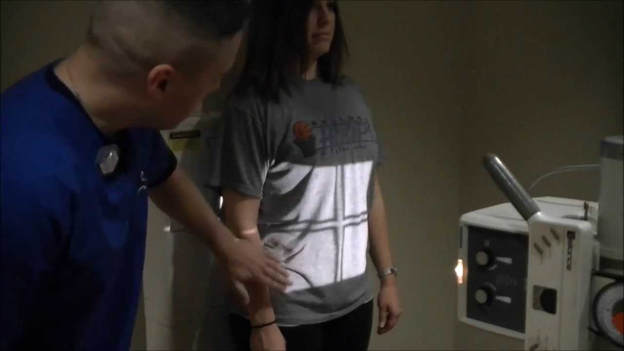
The standard abdominal radiograph (AXR) taken is a supine projection: x rays are passed from front to back (anteroposte-rior projection) of a patient lying down on his or her back. In some circumstances an erect AXR is requested: its advantage over a supine film is the visualisation of air-fluid lev-els. A decubitus film (patient lying on his or

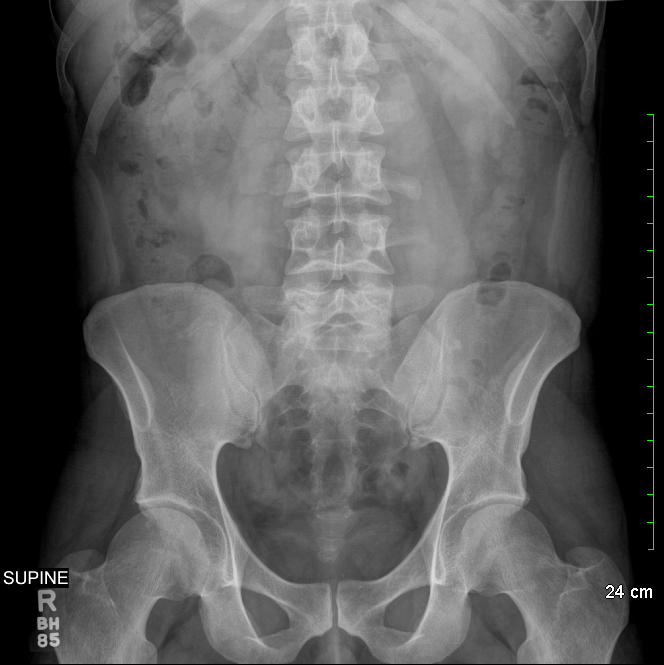
Supine Abdomen X Ray Anatomy
This present issue of Journal of Medical Radiation Sciences features a study by Geng et al assessing the usefulness of the erect abdominal radiograph, in addition to a supine radiograph, in the diagnostic work‐up of adult patients presenting with suspected intestinal obstruction.1 Conventional teaching stresses the identification of air‐fluid levels on erect radiographs as a feature of.

Xray abdomen supine showing dilated ascending and descending colon... Download Scientific Diagram
Plain abdominal radiography including supine and erect abdominal radiographs (SAR and EAR) is a frequently used image modality for preliminary evaluation of acute abdomen. We aimed to explore which one of the SAR or EAR has a higher diagnostic value in overall diagnosis of acute abdomen, including their respective advantages over each other for.

Abdomen supine AP view Xray K.U.B XRay Abdomen xray projection Part 1 By BL Kumawat
Supine (A) and erect (B) abdomen Xrays show dilated small bowel loops... Download Scientific
Abdominal radiographs are often the first diagnostic imaging tool for patients with acute abdominal pain. In most cases, a supine X-ray is sufficient,. Erect and supine abdomen radiographs are frequently obtained for patients presenting with acute abdominal pain (Gans et al., 2012). The actual cause of acute abdomen is frequently unknown.

PA supine abdomen Xray Diagram Quizlet
Background: Plain abdominal radiography including supine and erect abdominal radiographs (SAR and EAR) is a frequently used image modality for preliminary evaluation of acute abdomen. We aimed to explore which one of the SAR or EAR has a higher diagnostic value in overall diagnosis of acute abdomen, including their respective advantages over each other for the various underlying diseases.
Radiographic Imaging Supine and Erect Abdomen Projections YouTube
Abdominal radiographs are often the first diagnostic imaging tool for patients with acute abdominal pain. In most cases, a supine X-ray is sufficient, but in some cases, an erect abdominal radiograph may be warranted and can provide additional benefits. The aim of this study was to compare erect and.
Normal Abdominal X Ray / Normal Abdomen Xray Stock Photo Getty Images / Medgen uid the
The routine abdominal films consist of supine and upright views. If the patient cannot stand for an erect abdominal film and a PA view of the chest, the cross-table lateral projection with the right side elevated may be used to assess pneumoperitoneum and air-fluid levels. As little as 1 to 2 mL of free air in the peritoneal space may be.

Intestinal obstruction erect n supine abdominal x ray(2) YouTube
Introduction. Plain abdominal radiography (PAR) is often the initial diagnostic imaging tool for patients presenting with acute abdominal pain. 1 PAR in the acute setting may consist of supine and erect abdominal radiographs and an erect chest radiograph. 2, 3 The erect chest view is recommended for diagnosing chest pathologies, such as pneumonia, that mimic the symptoms of an acute abdomen. 4.

Xray Image Abdomen Supine Position Stock Photo 292611986 Shutterstock
Citation, DOI, disclosures and article data. The PA erect abdominal radiograph is often obtained in conjunction with the AP supine abdominal view in the acute abdominal series of radiographs. The erect abdominal radiograph has virtually disappeared from clinical practice in the United Kingdom, with studies dating back to the 1980s affirming.

xmlinkhub
Abdomen X-ray Guideline. Routine: 3 views • PA Chest • UPRIGHT PA or AP Abdomen (Include both hemidiaphragms) • SUPINE Abdomen (Include symphysis pubis). Only the right diaphragm must be included. • If unable to roll patient, do cross-table lateral abdomen with patient supine (May be utilized on rare occasions). Routine: 2.

Xray abdomen erect in (A) and supine in (B) showing multiple... Download Scientific Diagram
The standard abdominal radiograph is a supine projection: x-rays are passed from front to back (anteroposterior projection) in a patient lying on his or her back ( Figure 1-1 ). In some circumstances, an abdominal radiograph taken with the patient erect is requested; its advantage over a supine film is the visualization of air/fluid levels.

Supine Xray abdomen showing multiple rod shaped radio opaque... Download Scientific Diagram
Abdominal radiographs are often the first diagnostic imaging tool for patients with acute abdominal pain. In most cases, a supine X-ray is sufficient, but in some cases, an erect abdominal.
Approach to the Abdominal xray (AXR) Undergraduate Diagnostic Imaging Fundamentals
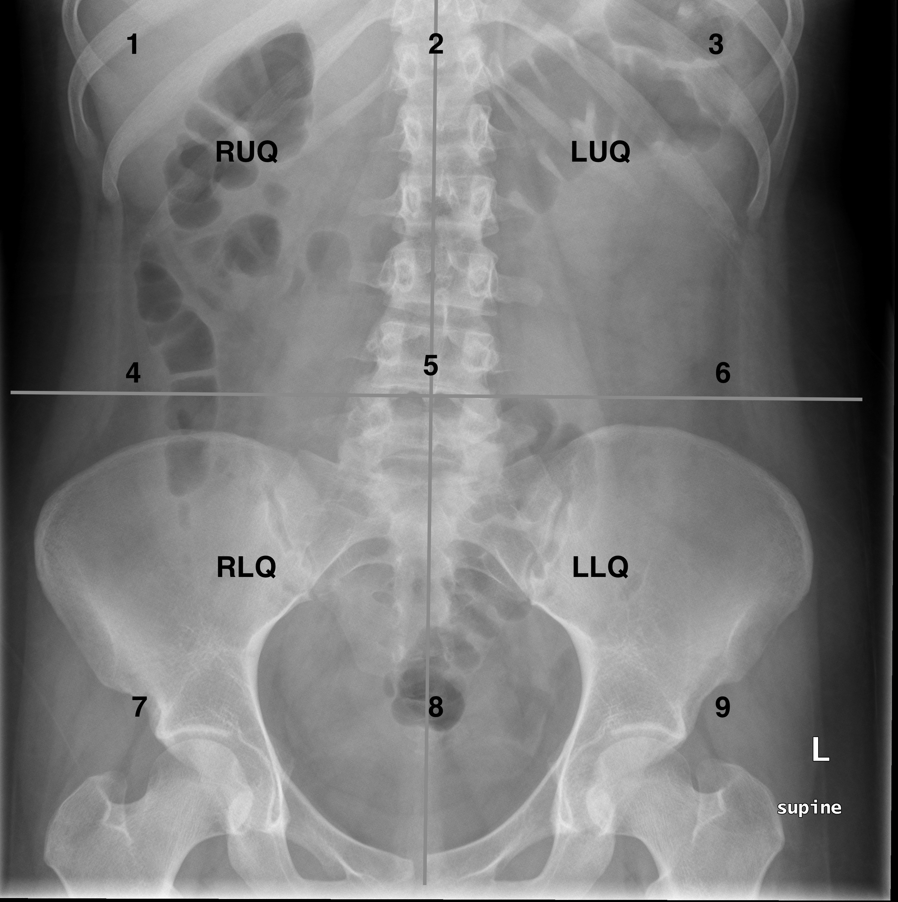
Bowel diameter. The upper limits for the normal diameter of different bowel segments are as follows: Small bowel: 3cm. Colon: 6 cm. Caecum: 9 cm. This is often referred to as the '3/6/9 rule'. A normal abdominal X-ray showing large bowel (white arrow) framing the small bowel (black arrow) 5. Example of faeces and its typical mottled.
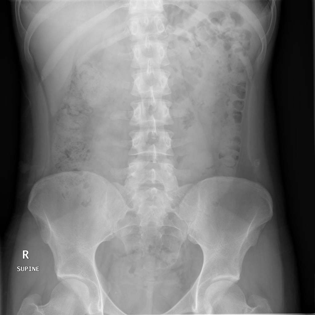
Normal Abdominal X Ray
Acute abdominal series is a set of abdominal radiographs obtained to evaluate bowel gas. The usual projections for this series are AP supine view (to estimate the amount of bowel gas or possible distension), PA erect view (to assess air-fluid levels), and PA erect chest radiograph (to rule out free air) . If the patient cannot tolerate upright.

Plain radiograph, supine (A) and erect (B), of a patient with acute... Download Scientific Diagram
Technique: The abdominal radiograph (AXR) is performed almost exclusively in the supine position and in the AP (anteroposterior) projection, i.e. the x-ray beam passes through the patient from front to back. Historically the abdominal radiograph was performed in both supine and erect postures, but this practice has been discontinued due to.