
Simple Labeled Human Respiratory System Diagram greeneyesstyle
The upper respiratory system, or upper respiratory tract, consists of the nose and nasal cavity, the pharynx, and the larynx. These structures allow us to breathe and speak. They warm and clean the air we inhale: mucous membranes lining upper respiratory structures trap some foreign particles, including smoke and other pollutants, before the.

System Label Respiration System To Label Human Anatomy Library Respiratory system anatomy
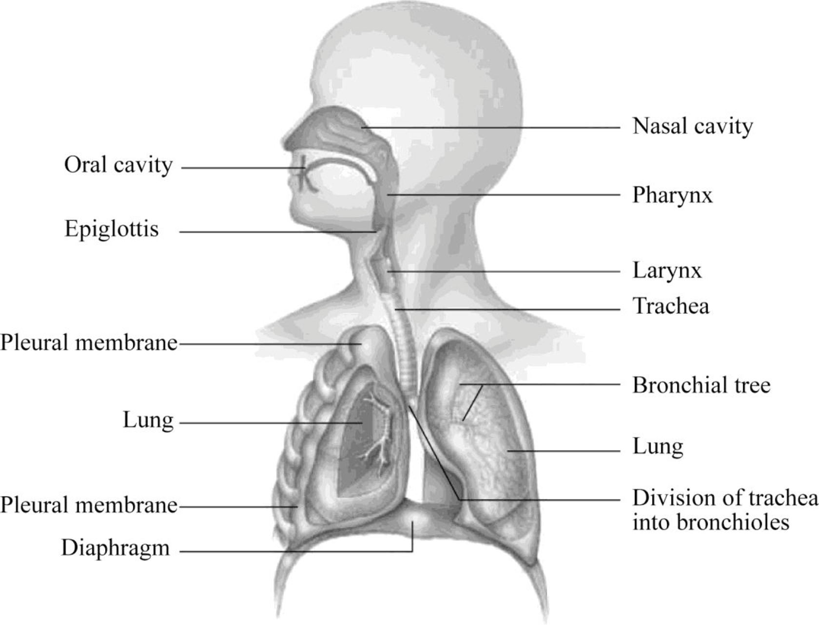
Portions of the respiratory system are also used for non-vital functions, such as sensing odors, speech production, and for straining, such as during childbirth or coughing (Figure 22.2). Figure 22.2 Major Respiratory Structures The major respiratory structures span the nasal cavity to the diaphragm.
Respiratory system HSC PDHPE
Diagram of the Human Respiratory System (Infographic) The primary organs of the respiratory system are the lungs, which function to take in oxygen and expel carbon dioxide as we breathe. The gas.

Human Respiratory System 7 Download Scientific Diagram
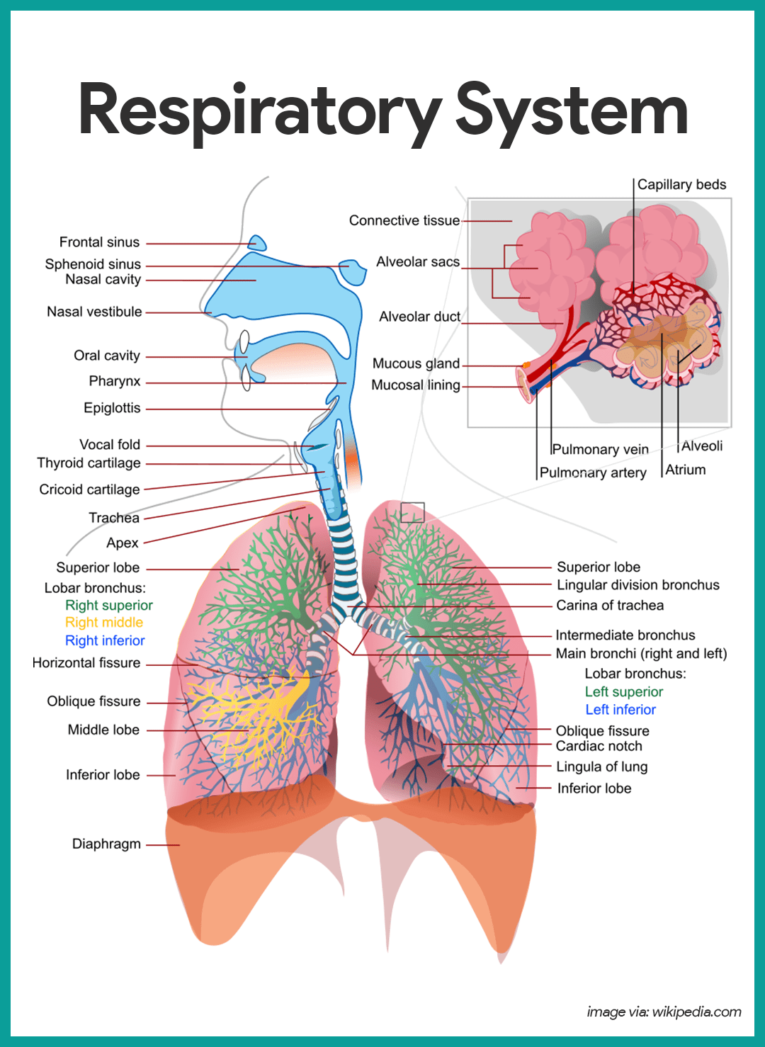
The nasal epithelium (Figure 20.6.1 20.6. 1 is lined with ciliated pseudostratified epithelial with goblet cells and this makes up the mucosal layer. Deep to this layer will be numerous bipolar cell nuclei. You will also find Bowman's (olfactory) glands that secrete mucus to help lubricate the mucosal layer and to dissolve odorants.
Respiratory System Diagram With Labels
Respiratory conditions. Summary. The respiratory system allows air to reach the lungs, from which oxygen enters the blood and circulates to all body cells. This system also removes waste gases.

Lung Diagrams 101 Diagrams
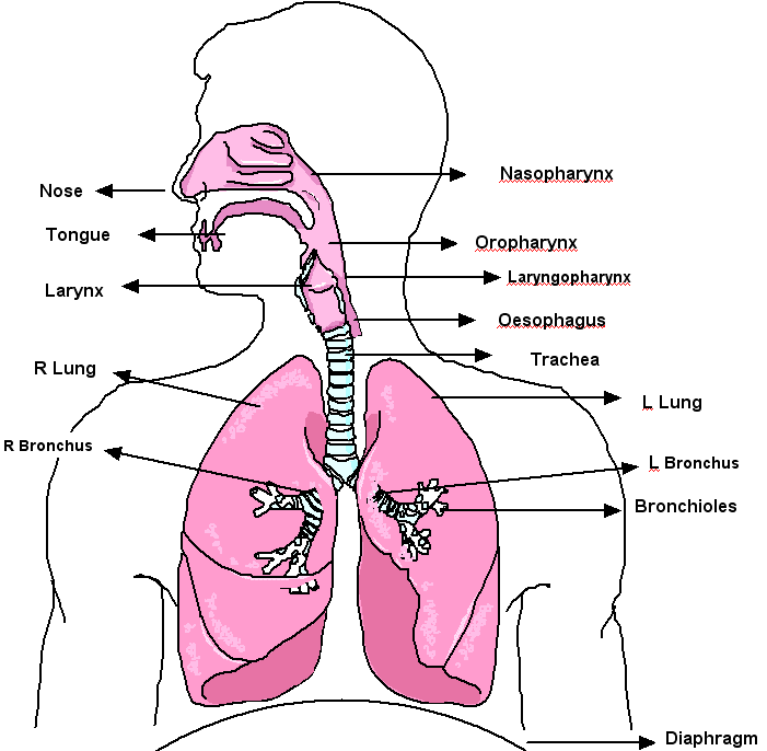
The major organs in the respiratory system are labeled. [Return to Figure 11.1]. Figure 11.2 image description: This figure shows a cross section view of the nose and throat. The major parts are labeled.. Diagram of the lungs with the major parts labeled (from top, clockwise): trachea, superior lobe, main bronchus, lobar bronchus,.

Schematic representation of the respiratory system. Download Scientific Diagram
The cells of the human body require a constant stream of oxygen to stay alive. The respiratory system provides oxygen to the body's cells while removing carbon dioxide, a waste product that can be lethal if allowed to accumulate. There are 3 major parts of the respiratory system: the airway, the lungs, and the muscles of respiration.
Respiratory System Anatomy and Physiology Nurseslabs
The respiratory system helps in breathing (also known as pulmonary ventilation.) The air inhaled through the nose moves through the pharynx, larynx, trachea and into the lungs. The air is exhaled back through the same pathway. Changes in the volume and pressure in the lungs aid in pulmonary ventilation.

Original file (SVG file, nominally 800 × 900 pixels, file size 330 KB)
The lungs are the main part of your respiratory system. Here is how lungs work as the center of your breathing, the path a full breath takes in your body, and a 3-D model of lung anatomy.
Standard Note Human Respiratory System
The respiratory system aids the body in the exchange of gases between the air and blood, and between the blood and the body's billions of cells. It includes air passages, pulmonary vessels, the.
Labeled Diagram Of The Respiratory System Of A Human
The respiratory system, also called the pulmonary system, consists of several organs that function as a whole to oxygenate the body through the process of respiration (breathing). This process involves inhaling air and conducting it to the lungs where gas exchange occurs, in which oxygen is extracted from the air, and carbon dioxide expelled.
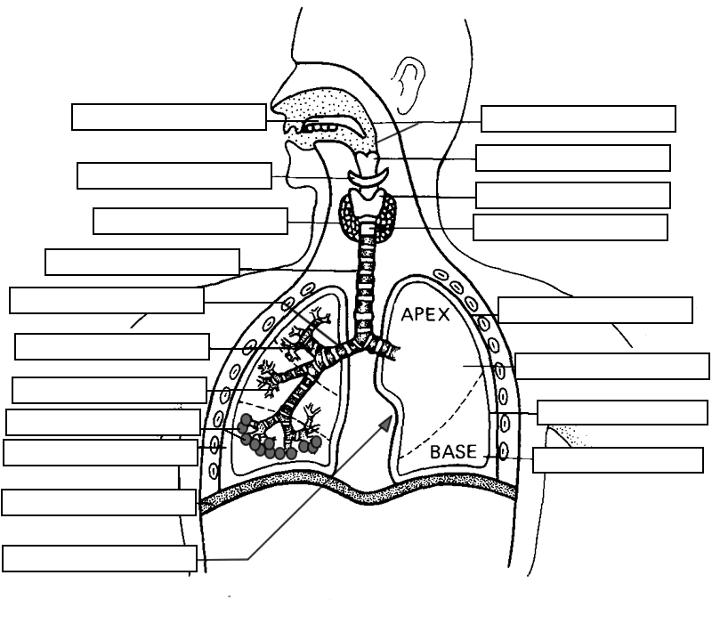
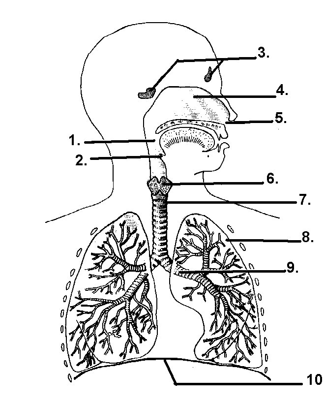
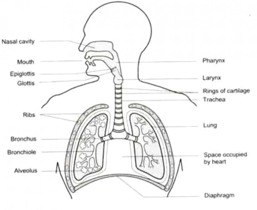
Label the structures of the Respiratory System
The Respiratory System (Label Diagram) Matching Quiz. Match each pair by dragging from rifgt to left. When complete choose the Check button. Check.

THE RESPIRATORY SYSTEM Resources that may be used
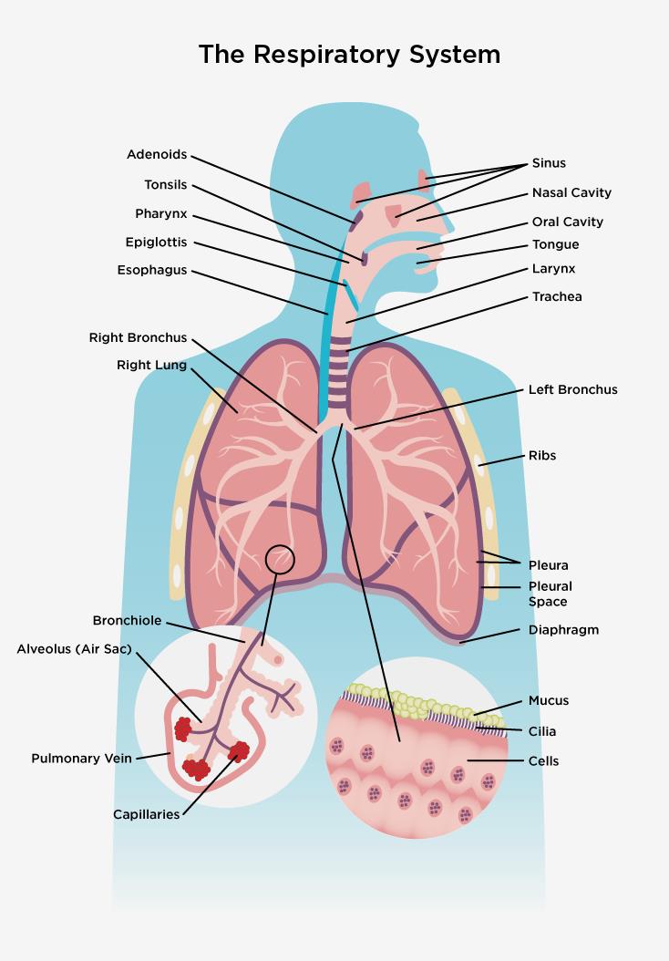
Respiratory System. Your respiratory system is made up of your lungs, airways (trachea, bronchi and bronchioles), diaphragm, voice box, throat, nose and mouth. Its main function is to breathe in oxygen and breathe out carbon dioxide. It also helps protect you from harmful particles and germs and allows you to smell and speak.

Introduction to Respiratory system Nursing Lecture
With a labeled diagram, you can see all of the main structures of an organ system together on one page - great for helping you to memorise the appearance of several structures and their relations. Unlabeled diagrams can then help you to put your memory to the test. Below, you'll find the respiratory system labeled and unlabeled on two.

Respiratory System Function & Organs Respiratory System Diagram
The respiratory system is how we bring oxygen into our bodies so that it can be absorbed into the blood, and how we get carbon dioxide out of our blood, so that it can be exhaled from the body. Its key organs are. the lungs, which provide huge surface area for absorbing gases. The series of hollow tubes (such as the trachea and bronchial tubes.
Respiratory system Canadian Lung Association
The respiratory tract conveys air from the mouth and nose to the lungs, where oxygen and carbon dioxide are exchanged between the alveoli and the capillaries. Sagittal view of the human nasal cavity. The human gas-exchanging organ, the lung, is located in the thorax, where its delicate tissues are protected by the bony and muscular thoracic cage.