
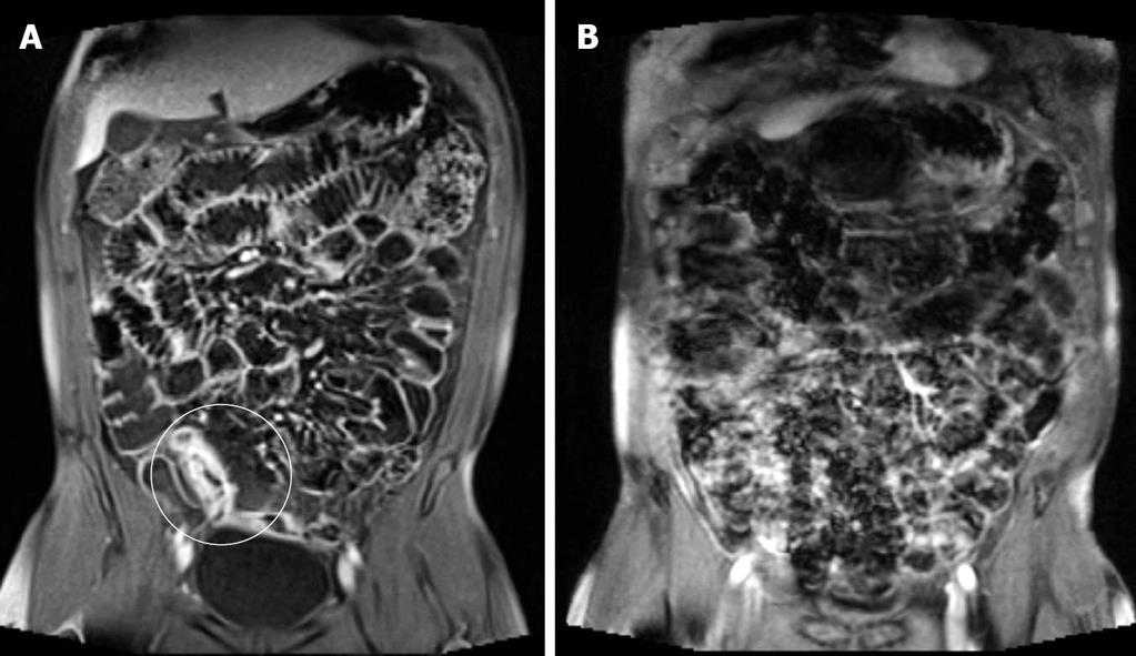
Representative small bowel MRI sequences. [A] TRUFIsequence small... Download Scientific Diagram
The side effects of MRI prep. "May have a slight laxative effect but will soon pass" said the cheery-sounding official hospital letter I received. Clearly, the person who wrote it did not have inflammatory bowel disease and was not aware of the actual definition of "slight." "Slight" is not how I would describe the laxative effect of this liquid.

Cine MR imaging of the small intestine in a case of bowel MRI YouTube
MRI Small Bowel Study This leaflet explains about MRI Small bowel studies, including the benefits, risks and any alternatives and what you can expect when you come to hospital. If you have any further questions, please speak to a doctor or nurse caring for you. What is an MRI scan? Magnetic Resonance Imaging (MRI) is a type of scan that uses.
Small bowel MRI enteroclysis or follow through Which is optimal?
MRI of the gastrointestinal tract is gaining clinical acceptance and is increasingly used to evaluate patients with suspected small-bowel diseases. MRI may be performed with enterography or enteroclysis, both of which combine the advantages of cross-sectional imaging with those of conventional enteroclysis. In this paper, MRI features of primary small-bowel neoplasms, the most important signs.

MRI to evaluate small bowel Crohn's disease
In this article we will discuss the MRI-features used to evaluate Crohn's disease of the small bowel and the colon. A systematic approach is presented to grade disease activity resulting in a simple classification of mild, moderate and severe disease. This is sufficient for most therapeutic decisions. Introduction.
The Radiology Assistant Small Bowel Tumors
MRI techniques available to evaluate the small bowel include MR enterography (MRE) and MR enteroclysis. In MR enteroclysis, enteric contrast is administered directly via a nasoenteric tube, providing superior distention of small-bowel loops compared to oral ingestion in MR enterography. However, MR enteroclysis remains of limited availability.

Small Bowel Obstruction What to Look For RadioGraphics
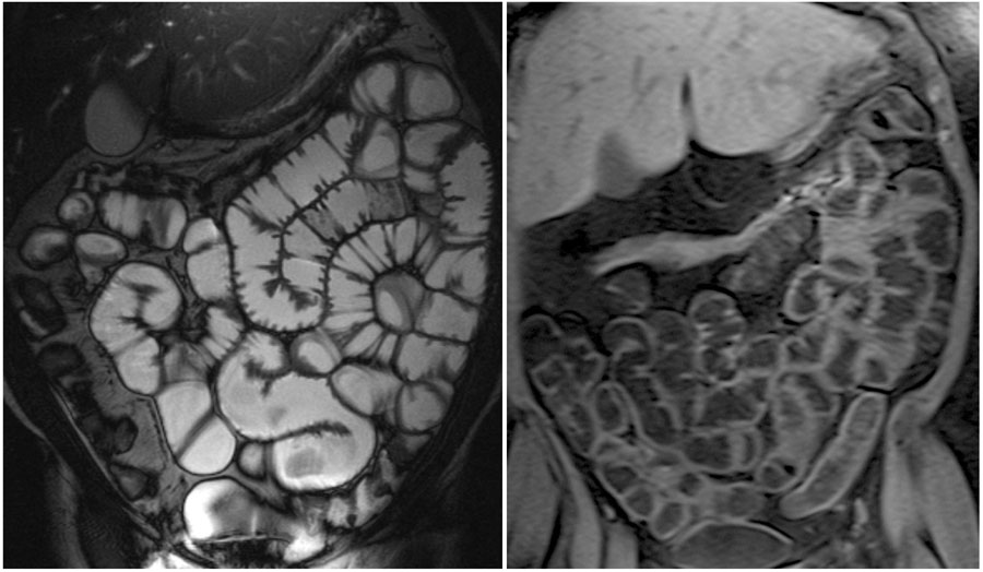
Imaging of the small bowel is most often performed on 1.5-T MRI, although the examinations can be performed on 3-T MRI with slight adaptation of the sequences. All sequences should be performed in breath-holds. The patient's position in the MRI can be supine or prone.

Small Bowel Obstruction due to Adhesions Small Bowel Case Studies CTisus CT Scanning
Axial contrast-enhanced CT enterographic image shows a short-segment stricture of the ileum (arrows). The radiologist should be aware of the spectrum and typical imaging appearances of small bowel diseases to accurately guide clinical management. Disclosures of conflicts of interest.—D.H.B. Editorial board member of RadioGraphics.

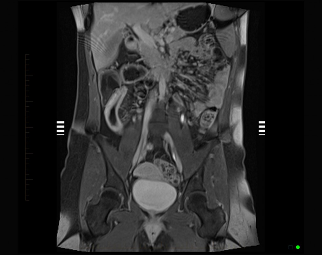
A coronal view of the patient's abdomen on MRIsmall bowel... Download Scientific Diagram
During your MRI scan. You will be asked to drink between 800 to 1500mls of the Mannitol sugar solution after you have had safety checks with a radiographer or radiology assistant. This solution should reach the end of your small bowel within 40 to 60 minutes. Sometimes this can take longer. The MRI scan can then begin.
MRI to evaluate small bowel Crohn's disease
MRI Scanning for Small Bowel Disease Introduction This leaflet tells you about having an MRI scan for small bowel disease. The small bowel is difficult for doctors to get to with an endoscope (camera). It lies beyond the stomach and duodenum, and loops around for several metres to the right lower abdomen where it joins the large bowel.

CT of Small Bowel GIST Tumors Imaging and Theory Part 2 YouTube
MRI Small Bowel Imaging Page 5 of 8 order to provide a full and comprehensive report on your scan. A cannula (small needle) will be inserted into a vein in the arm or hand before the scan. The first is a muscle relaxant (Buscopan) used to settle bowel movement obscuring the area
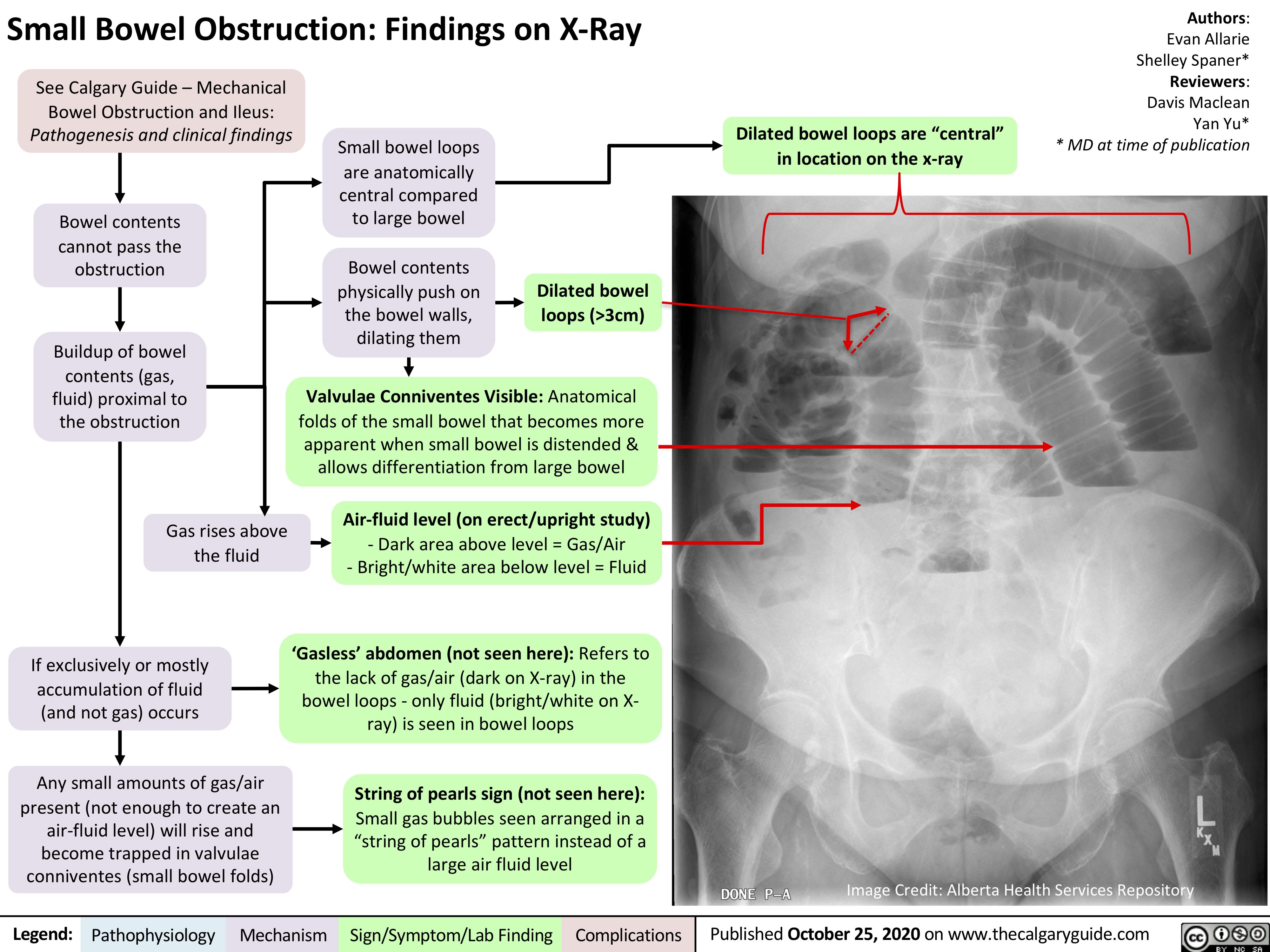
Small Bowel Obstruction Findings on XRay Calgary Guide
Ref: B-264/Imaging/MW/MRI of the small bowel v4. PDF: MRI of the small bowel [pdf] 181KB . YOU MUST ATTEND YOUR APPOINTMENT 1 HOUR PRIOR TO YOUR APPOINTMENT TIME . Failure to do so may mean your scan will be postponed. What is MRI (Magnetic Resonance Imaging) of the small bowel? This is a MRI examination of the small bowel and abdomen.

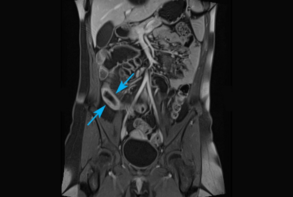
Abdomen MRI shows inflamed distal small intestine (terminal ilium) in patient with
Magnetic resonance small bowel is an imaging test that lets your doctor see detailed pictures of your small intestine. It can help identify inflammation, bleeding, and other problems. The test uses magnets and sound to create detailed images of your organs. Before the test, oral contrast dye is given to highlight the small intestine.

Bowel pathology Radiology Cafe
It's easy to find out more about treatment by giving us a call or completing our enquiry form. 01580 363158. Make an enquiry. Small bowel MRI or MR enterography uses an MRI scanner to produce detailed images of your small intestine to help identify small bowel conditions.
MRI to evaluate small bowel Crohn's disease
The purpose of utilizing an oral contrast agent is to achieve specific signal intensities in the bowel lumen on different types of MRI images. On T2-weighted images, the aim is to produce a high signal intensity in the bowel lumen, while on T1-weighted images, a low signal intensity in the lumen is desired.

Small Bowel Crohn Disease at CT and MR Enterography Imaging Atlas and Glossary of Terms
MR enterography ( MRE ), also known as MRI small bowel study, is a non-invasive technique for the diagnosis of small bowel disorders. Note: This article is intended to outline some general principles of protocol design. The specifics will vary depending on MRI hardware and software, radiologist's and referrer's preference, institutional.
Radiological Anatomy Small Intestine Stepwards
Representatives from the Society of Abdominal Radiology Crohn's Disease-Focused Panel, the Society for Pediatric Radiology, the American Gastroenterological Association, and other international experts recently reported consensus recommendations for standardized nomenclature for the interpretation and reporting of CT enterography and MR enterography findings of small bowel Crohn disease.
- Moroccan Spice Mix Crossword Clue
- S S Lazio Vs As Roma Lineups
- Hotels Near To St Pancras Station London
- Four Points By Sheraton Seoul
- Jujutsu Kaisen Dubbed Season 2
- Grain Harvest Season Western Australia
- Red Bellied Black Snake Size
- What S In The Box Today
- Man City Vs Liverpool F C Stats
- Crown Of Thorns Star Fish