
Melanoma On Lip Photos
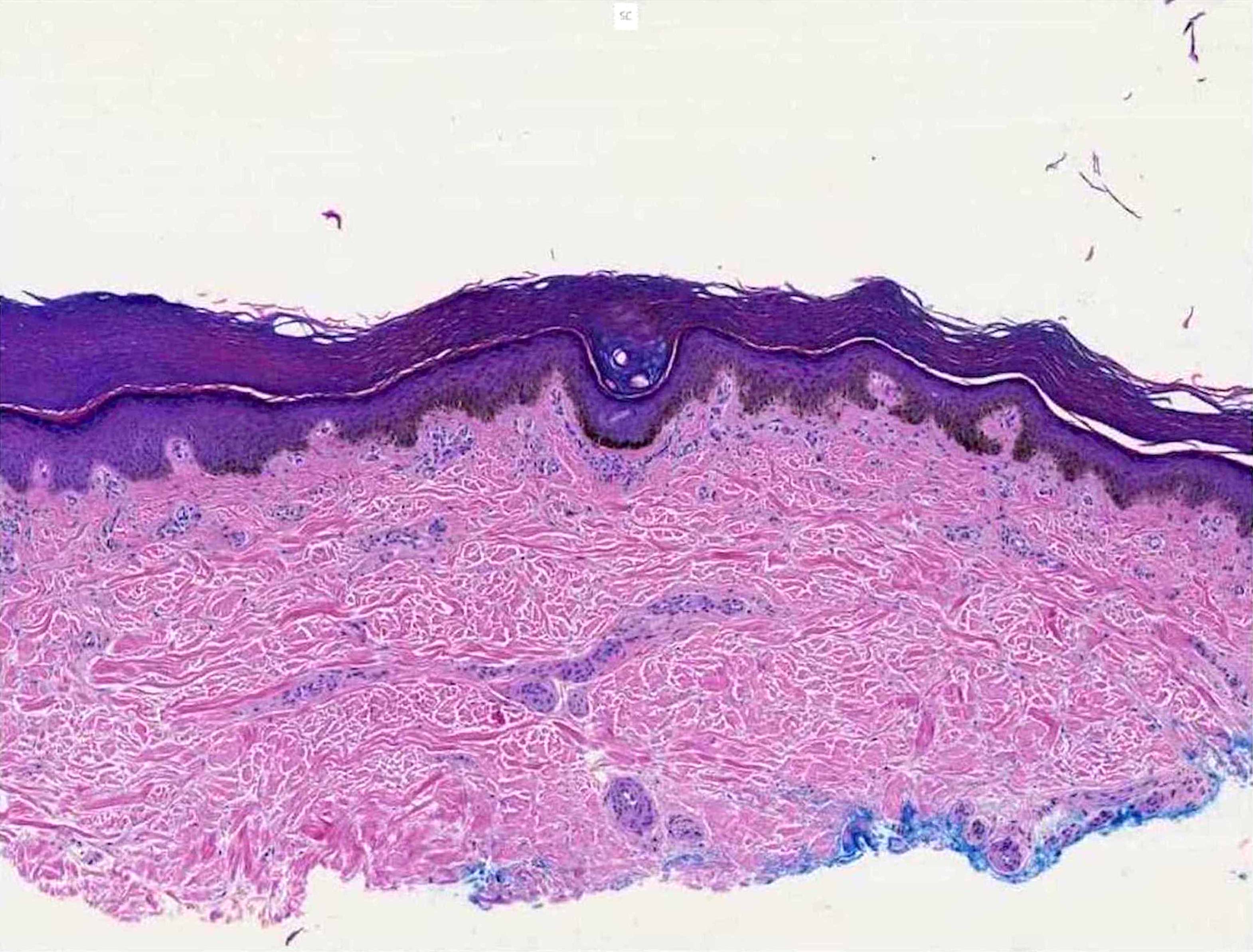
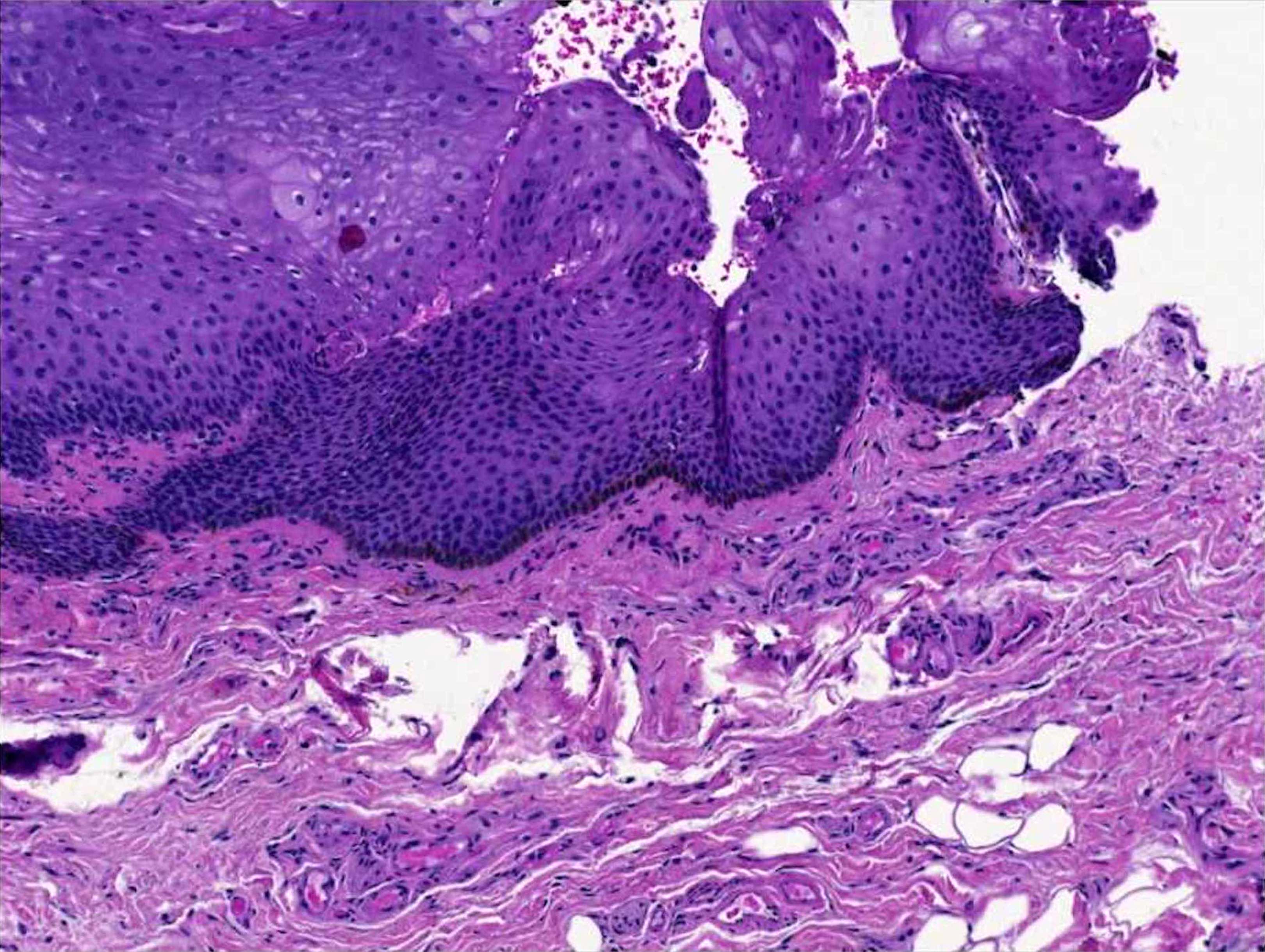
The oral melanotic macule is a small, well-circumscribed, brown-to-black that occurs commonly on the lips and gingiva, followed by the palate and buccal mucosa. Labial melanotic macule. B). Increased numbers of me-lanocytes along the junctional zone (H&E, x 200). C) Intraoral me-lanocytic nevus located on the left buccal mucosa.

Mucosal melanocytic macules
Lip melanosis is a cosmetically disfiguring condition frequently encountered in the Dermatology outpatient department. Lip melanosis has multifactorial etiology.. 2.1 sessions in labial melanotic macule, 3 sessions in post-inflammatory hyperpigmentation, and 4.2 sessions in diffuse pigmentation. Recurrence was seen in two patients. The only.
Pathology Outlines Melanotic macule
Excerpt from my virtual slide session for the Stony Brook University dermatology residents on 11/18/21. Full video available here (FREE): https://kikoxp.com/.

Hyperpigmented macules on the patient's lips. Download Scientific Diagram
The term labial melanotic macule of the lips (MML) is used to describe a flat-pigmented lesion on the vermilion border, with an estimated prevalence of up to 3% of the population. Most MMLs are located on the lower lip and are more common in female patients. MMLs are mostly associated with hyperpigmentation of the basal keratinocytes with normal to a slightly increased density of melanocytes.

Hyperpigmented lip lesions differntial diagnosis Dermatology Games
Labial and oral melanotic macules are commonly encountered in a broad range of conditions ranging from physiologic pigmentation to a sign of an underlying life-threatening disease. Although Laugier-Hunziker syndrome (LHS) shares some features of labial and oral pigmentation with a variety of conditions, it is a benign and acquired condition, frequently associated with longitudinal melanonychia.

Multiple small, dark brown macules on the lip vermillion and perioral... Download Scientific
Signs & Symptoms. The most common locations for an oral melanotic macule include the: Lips, especially the lower lip. Gums (gingiva). Inner cheek (buccal mucosa). Roof of the mouth (hard or soft palate). An oral melanotic macule appears as a brown or grayish-brown macule (a small flat, smooth area of skin or mucosa) usually less than 7 mm in.
Lip Pigmentation Treatment in Singapore Dr Wong Soon Tee
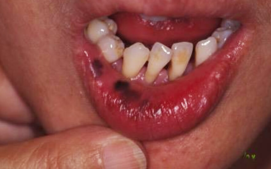
Fifteen patients (9 female and 6 male, aged 5-65 years) with oral melanotic macules, all located on the vermilion borders of the lips, were treated with simple cryosurgery. In 12 of the patients, the melanotic macule was a solitary lesion. Three patients had multiple lesions (Fig 1, A).
Orale melanosis
Benign melanotic macules (MAC) are the most frequent cause of lip pigmentation and can usually be distinguished from melanoma (MEL) using clinical and dermoscopic criteria. 1-4 However, the diagnosis of MAC can be challenging, and close follow-up and/or a lip biopsy may be needed to establish a diagnosis with confidence. Biopsying lip lesions is troublesome for patients because it is often.
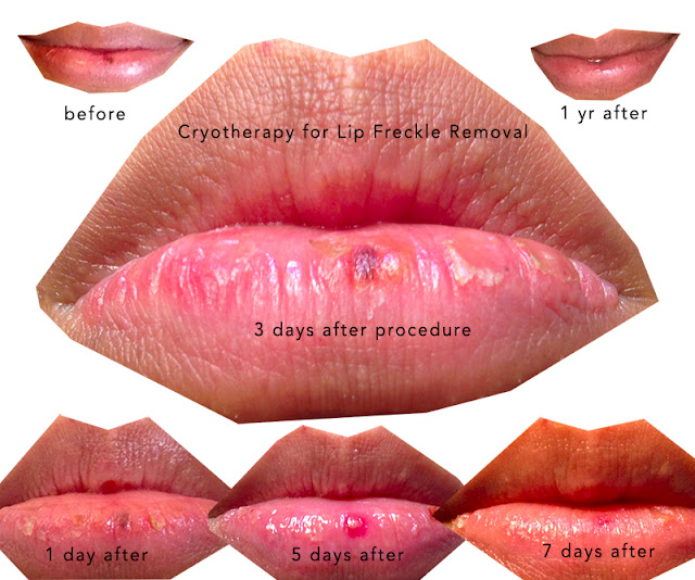
My Lip Freckle Removal Story
A mucosal melanocytic macule is a well-defined, oval, brown to black, flat patch, usually found on the lip (a labial melanocytic macule). Lesions can also be found in the mouth (oral melanotic macule), on the vulva (vulval labial melanotic macule; vulval melanosis) or on the penis (penile melanotic macule).
Pathology Outlines Melanotic macule
Melanotic macule. Melanotic macules are the most common oral mucosal lesions of melanocytic origin. 3 These small, solitary, well-circumscribed and often uniformly pigmented lesions develop most commonly in adult female patients. Any mucosal site may be affected but the lower lip, gingiva and palate are the most common areas.
oral melanotic macule pictures, photos
An oral melanotic macule is a benign hyperpigmented macule that may be found on the lips or oral mucosa in approximately 3% of the general population. It is caused by an increase in focal melanin deposition that is occasionally associated with an increased number of melanocytes. Melanotic macules are most commonly found on the vermillion border.

Discrete brown black macules on the lower lip. Download Scientific Diagram
Labial melanotic macule was described by Weathers et al. (1) and is a clinically and histologically distinctive benign pigmentary abnormality of the lip (1-4, 10-12). The majority of patients are young adult women with a mean age at onset of approximately 30 years. Labial melanotic macule typically appears on the vermilion border of the.

Pigmented Lesions of the Oral Mucosa Pocket Dentistry
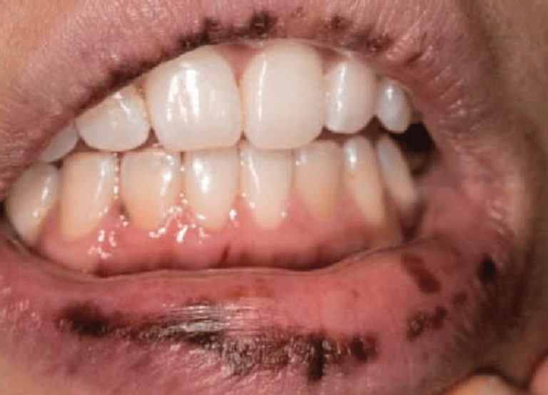
Labial macules occur most frequently in women who are fair-skinned and have had ample sun exposure. They characteristically present in the third decade of life and appear as well-defined dark brown to black macules on the lower lip, ranging in size from 2 to 6 mm. The condition was first reported in 1976, 1 and one study noted an incidence of 3.

(A)(C) Multiple discrete dark brown macules over lips, labial mucosa,... Download Scientific
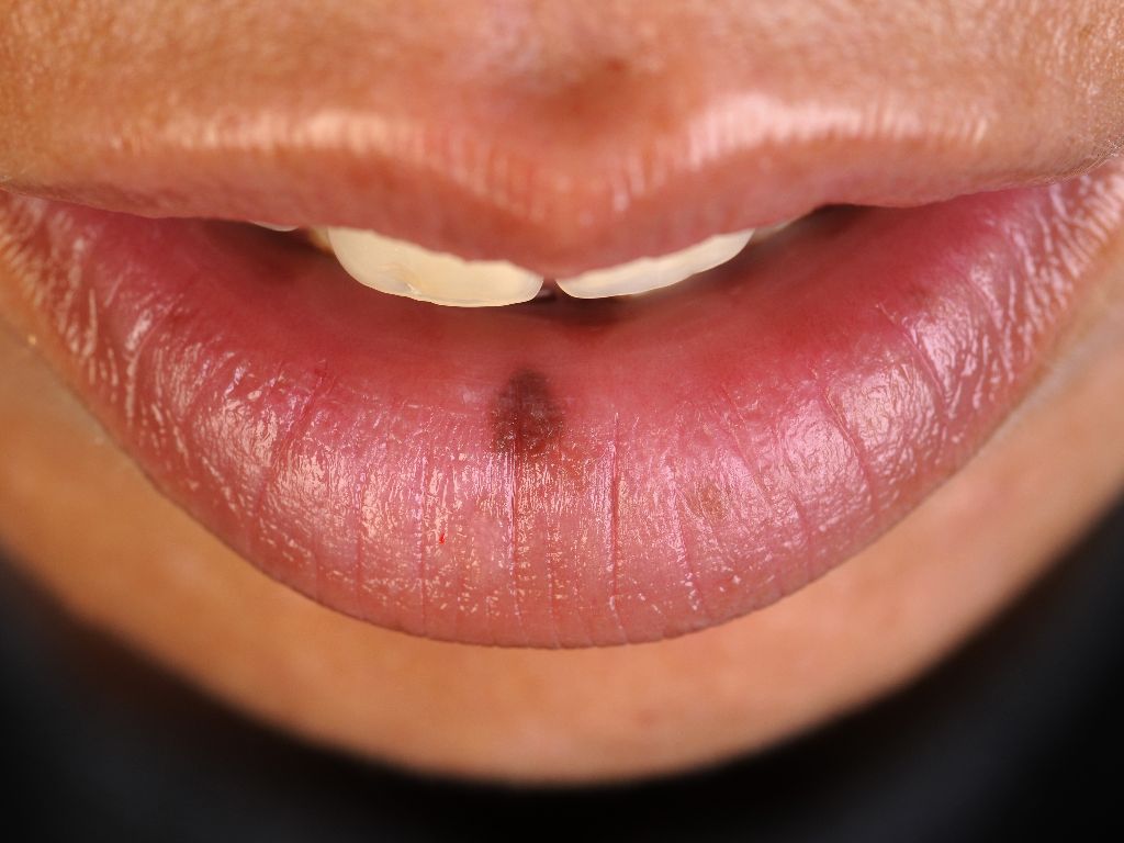
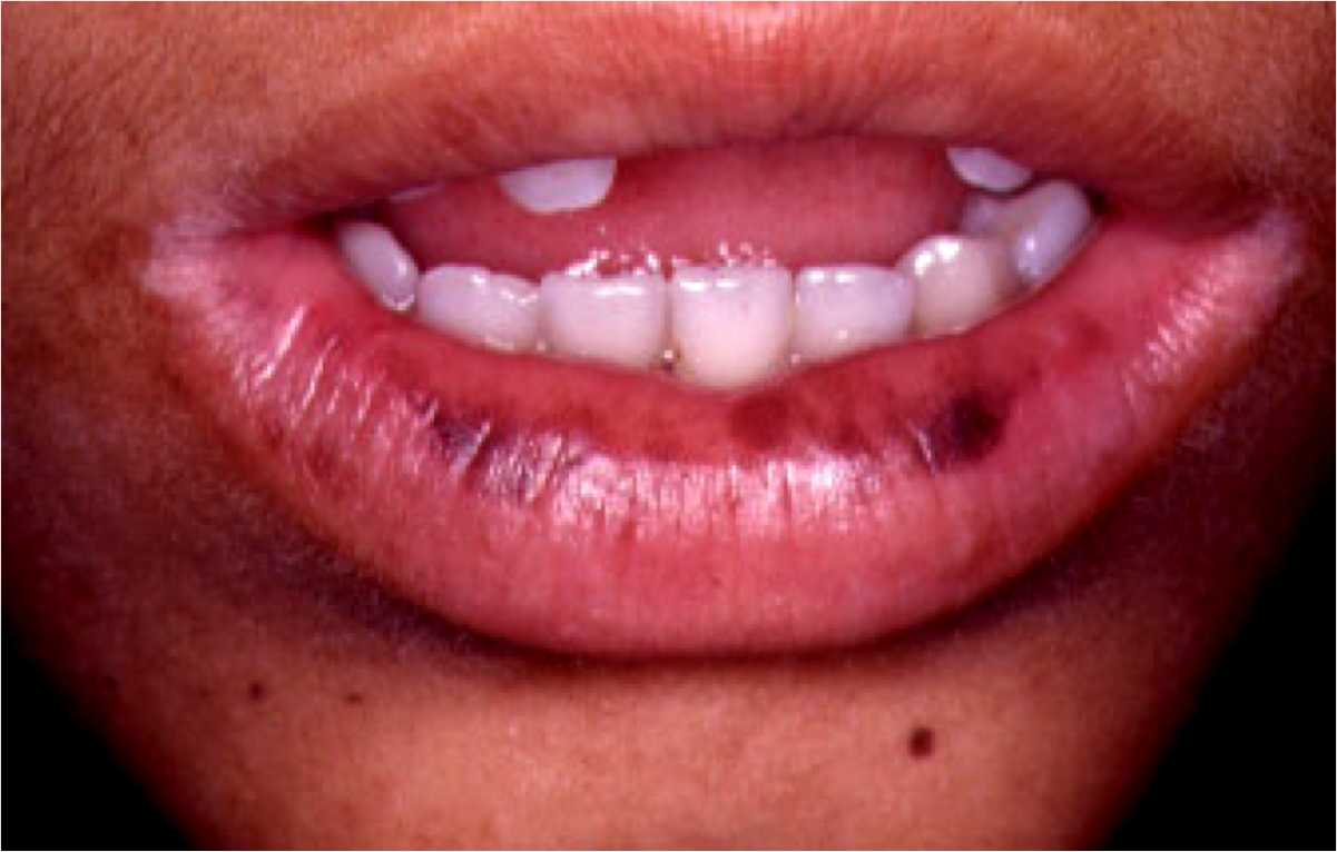
A labial melanotic macule is a well-defined, oval, brown to black, flat patch on the central third of the lower lip. It is the name for a freckle arising on the lip. It is also sometimes called a labial lentigo and when multiple lesions are present, mucosal melanosis.
Hyperpigmented lip lesions differntial diagnosis Dermatology Games
A macule indicates the presence of a flat, distinct, discolored area of skin less than 1 cm wide. A macule usually does not involve any change in the thickness or texture of the affected skin. Labial Melanotic Macules are observed on the lips, especially in the lower lip. It is a commonly observed skin condition in children and adults.

Lower lip showing smoker's melanosis. 2 Download Scientific Diagram
The Labial Melanotic Macule. From the Division of Dermatology, University of Louisville School of Medicine. • A series of 41 cases of melanotic lesions of the lip from 1980 to 1984 are reported. These lesions occur preponderantly in young white women on the lower lip. The term labial melanotic macule is suggested for these lesions.